Improved Method for Treatment of Burns

The use of meshed split skin autographs (SSGs) combined with autologous cultured proliferating epidermal cells provided better wound healing and less scarring compared to using SSGs alone.

In a 40-patient clinical trial, researchers from The Netherlands found that such technique provided better results for patients who suffered serious and deep burns. The usual method of treating burns was the use of split skin autographs.

The new technique developed by the researchers used autologous (self-donated), cultured proliferating epidermal cells that were “harvested” from a small donor site, and “seeded” in a collagen carrier that could, in theory, enhance the wound healing rate and improve scar quality.

Dr. Shinn-Zong Lin, Vice-Superintendent for the Center of Neuropsychiatry, professor of Neurosurgery at China Medical University Hospital, and coeditor-in-chief for Cell Transplantationwas quoted saying that this study offers a promising, improved therapeutic method for treating severe burns.

According to Dr. Esther Middelkoop of the VU University Medical Center in Amsterdam, a co-author of the study,

The rate of epithelialization in the experimental treatment was statistically significantly better when compared to the standard treatment. We also established improved pigmentation for the wounds treated with cultured ECs. Scar quality impacts patients’ lives in many ways due to cosmetic and functional concerns.

Additionally, there is a high economic burden on patients due to extended hospital stays and the cost of rehabilitation and reconstructive therapies. Because of this, additional research in burn wound treatment and the improvement of scar quality will always be of the highest priority.

It was revealed that epithelial cells applied to a carrier system could, in fact, reduce wound healing time and improve both short-term and long-term functional as well as cosmetic scar quality.

It was observed that the primary outcome was wound closure after five to seven days, said the researchers. Secondary outcomes were safety and scar quality, which were measured at three and 12 months.

Read more on: http://ingentaconnect.com/content/cog/ct/pre-prints/content-CT-1380_Gardien_et_al

Have Researchers Found the Best Method of Harvesting Autologous Fat?

A study published in the Journal of the American Society of Plastic Surgeons sought to examine the method that will optimize the process of harvesting lipoaspirate before grafting.

Dr. Emily Cleveland, together with her colleagues from the New York University Institute of Reconstructive Plastic Surgery, examined various articles on the methods of processing human fat for autologous grafting in an evidence-based review.

They found that there was no single viable method that may be advocated as the best technique for lipoaspirate process.

Autologous fat harvested through liposuction techniques is used by both cosmetic and reconstructive surgeons. As a filler, it has several advantages including availability, biocompatibilty, ease of harvest without risk of allergic reaction or rejection, and it's inexpensive.

This technique has been used in several procedures including, but not limited to, facial rejuvenation, breast augmentation and reconstruction, treatment of congenital anomalies, and improvement of soft-tissue damage due to radiation therapy.

Despite its advantages, the autologous fat transfer technique is also wrought with setbacks. Viability and the retention of fat graft cannot be predicted with certainty because of lack of clear data pinpointing factors which may be responsible for the variability in results.

However, there is a wide belief among practitioners that the lack of standardized procedure, especially with that of postharvest fat processing, significantly contributes to the variability. Currently, several techniques of fat harvest before transplantation are used.

Among them are the use of simple decantation, cotton gauze rolling, centrifugation, and washing in physiologic solutions.

In the study, randomized controlled trials, clinical trials, and comparative studies comparing at least two of the following techniques were included: decanting, cotton gauze (Telfa) rolling, centrifugation, washing, filtration, and stromal vascular fraction isolation.

Results of the study were as follows:

  • There is a lack of superior method for processing harvested lipoaspirate.
  • Simple decantation has previously been demonstrated to preserve a large number of intact and nucleated adipocytes. However, it allows a significantly greater amount of aqueous and lipid contaminants to remain in the specimen, particularly hematogenous cells and other materials that are believed to be proinflammatory and thus harmful to graft survival. Recent publications further confirm this, demonstrating lower rates of decanted graft viability relative to centrifuged and washed specimens.
  • There are limited data to suggest that cotton gauze rolling of the lipoaspirate produces a graft largely free of contaminants, with superior in vitro adipose-derived mesenchymal stem cell content and high rates of in vivo retention but the technique is quite labor intensive.
  • Centrifugation is perhaps the most widely used technique for postharvest fat processing, and has previously been considered the criterion standard. The most commonly used settings are those described by Coleman, in which lipoaspirate is spun at 1200 g (3000 rpm) for 3 minutes, followed by discarding the aqueous inferior layer and wicking off the free oil top layer. The middle adipose layer is then grafted. Some have suggested this may not be the most viable technique, in that it fails to incorporate the “pellet,” which contains the highest number of adipose-derived mesenchymal stem cells in the harvested specimen. Recent literature has demonstrated lower rates of graft viability after centrifugation relative to washing,although equivalent or superior results have been shown by some after “soft” centrifugation (400 g for 1 minute). Nevertheless, other research continues to support the equal effectiveness of standard centrifugation in preserving adipose-derived mesenchymal stem cells and producing viable in vivo grafts.
  • Washing the lipoaspirate has previously been demonstrated to preserve both a large number of mesenchymal stem cells and a large number of adipocytes, thus satisfying both theories for graft survival. Several commercially available technologies that use washing techniques also appear promising for efficient, effective processing of lipoaspirate. This finding is somewhat confounded, however, by the use of multimodality technologies such as those used by Salinas et al. (washing then Telfa rolling or centrifugation), and processing with the Puregraft and Revolve systems, which first filter the lipoaspirate before washing. In addition, limited or no data are available to demonstrate in vivo superiority of these techniques.
  •  Filtration methods appear to eliminate contaminants, and maintain viable adipocytes and a large portion of adipose-derived mesenchymal stem cells. This processing technique may be more efficient in producing viable graft material for large-volume fat transfers, which are becoming increasingly popular among both cosmetic and reconstructive surgeons. The Tissu-Trans Filtron inline filtration system holds promise, but there are to date only extremely limited data available to support its use.
  • Similarly, there is only limited evidence to date to support the supplementation of processed lipoaspirate with additional stromal vascular fraction. Although viable isolation methods have been developed, a great deal of further research is required to determine whether this additional cost and effort is justified by superior clinical outcomes. The Celution 800/CRS System may be a viable method for isolating stromal vascular fraction in clinical settings for augmentation of autologous fat used for grafting; however, no subsequent in vivo study was performed to demonstrate its superiority relative to the other proprietary systems examined in this study. 

In conclusion, the authors said that they 

 did not find compelling evidence to advocate a single technique as the superior method for processing lipoaspirate in preparation for autologous fat grafting. A paucity of high-quality data continues to limit the clinician’s ability to determine the optimal method for purifying harvested adipose tissue. Novel automated technologies hold promise, particularly for large-volume fat grafting; however, extensive additional research is required to understand their true utility and efficiency in clinical settings.

More information about the study can be found at: http://journals.lww.com/plasreconsurg/Fulltext/2015/10000/Roll,_Spin,_Wash,_or_Filter__Processing_of.16.aspx?WT.mc_id=HPxADx20100319xMP

Viability of Autologous Fat Calf Grafting

Research shows that autologous calf fat grafting is a viable alternative to traditional implant-based calf augmentation for congenital calf discrepancies and aesthetic pseudo-varus deformity.

According to the study published in the official publication of the American Society for Aesthetic Plastic Surgery (ASAPS), Aesthetic Surgery Journal, fat grafting for slender calves provides results that are comparable to those obtainable via traditional methods. 

Some patients who want to undergo calf implants are concerned with the risks associated with the surgery and the possibility that strenuous exercise may cause the implant to move. Some plastic surgeons use silicone implants that is selected based on the patient's anatomy. These implants are placed under the fascia of the original calf muscle that is strong enough to withstand physical sporting activities.

Researchers Gerhard S. Mundinger and James E. Vogel pointed out in the research that there are few studies published regarding the advantages of fat grafting for calf augmentation and re-shaping compared with the traditional silicone calf implants.

According to Dr. James E. Vogel,

Autologous fat augmentation offers a number of advantages over calf implants, including liposuction in adjacent areas to improve calf contour, smaller incisions, additional augmentation through subsequent fat grafting, durable results, lack of foreign body reaction, and precise patient-specific adjustments not possible with off-the-shelf implants.

Medial and lateral calf augmentation was accomplished with injection of prepared autologous lipoaspirate intramuscularly and subcutaneously.

Thirteen patients underwent calf augmentation and reshaping with autologous fat grafting over a period of five years. Ten patients underwent bilateral calf augmentation, and three cases were performed for congenital leg discrepancies.

Prior to the fat transfer, local anesthesia was injected to utilize the smallest amount of effective anesthetic volume. This was also done to precisely place it into the muscle resulting in less sedation and more rapid postoperative recovery.

Fat was harvested from the abdomen, lateral thigh, medial thigh, waistline, flanks, axilla, upper back, and hips. Irrespective of the fat harvest site, liposuction was also performed at the knee to improve contour.

A mean of 157 cc of separated fat was transferred per leg, with roughly 60% and 40% transferred into the medial and lateral calf, respectively. Injections were first performed directly into the calf muscles and then into the subcutaneous calf tissue.

Four patients underwent a second round of autologous fat injection for further calf augmentation because they desired additional volume. Fat volume was judged to be sufficient when the calf was minimally firm but not tense. At a mean of 19.6 months of follow-up, durable augmentation in calf contour was documented by comparison of standardized preoperative and postoperative photographs.

The research concluded that the use of autologous fat transfer is a viable option compared to the use of traditional silicone implants.

Read more on: http://asj.oxfordjournals.org/content/early/2015/09/01/asj.sjv166

Scar Management Techniques for Surgeons

Each year, it is estimated that around 100 million people in developed countries acquire scars after undergoing elective surgery and surgery for trauma. In a purely cosmetic surgical procedure such as an aesthetic breast surgery, scarring is viewed as a source of dissatisfaction among patients.

Prevention and treatment of unaesthetic scar formation after an operation greatly rests on plastic surgeons who perform these operations. Scarring may have several unpleasant aesthetic and psychological consequences to the patients including diminished self esteem, stigmatization, anxiety and depression.
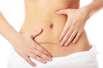
In a study published at Journal of Plastic, Reconstructive & Aesthetic Surgery, an international, multidisciplinary group of 24 experts developed a set of practical, evidence-based guidelines for the management of linear, hypertrophic and keloid scars which could be useful for surgeons, dermatologists, general practitioners and other physicians involved in the prevention and the treatment of scars.

Here are some of the results:

1. After a surgery, prevention of abnormal scar formation should be a priority. In an elective surgery, the position and length of scar is to be greatly considered. As much as possible, the incision should be parallel to the relaxed skin tension lines. During the operation, the surgeon should ensure that excessive tension on the wound edges is avoided. Several measures may also be done to reduce inflammation, provide rapid wound closure, reduce the risk of infection, and provide an early surgical wound coverage.

2. Following a wound closure, scar prevention consists of three phases: tension relief, hydration/taping/occlusion, and pressure garments. Recent studies show that offloading mechanical forces using a stress-shielding device made from silicone polymer sheets and pressure-sensitive adhesive significantly reduced scar formation. Also, the use of botulinum toxin A decreases tensile forces on post-surgical scars and results in significant improvements in the cosmetic appearances of scars compared with placebo injections.

3. Silicone products may help to prevent excessive scar formation by restoring the water barrier through occlusion and hydration of the stratum corneum and need to be used as soon as the wound/suture is healed. Moisturizing emollient and humectant creams and moisture-retentive dressings such as silicone sheets and fluid silicone gel have been shown to be beneficial for itching scars, and can also reduce the size and pain or discomfort associated with scars as well as improving their appearance.

4. Randomized studies in animals and humans have shown that ultraviolet radiation increases scar pigmentation and worsens their clinical appearance. A preventive measure of avoiding exposure to sunlight and the continued use of sunscreens with a high to maximum sun protection factor (>50) until the scar has matured is recommended.

5. For patients with linear hypertrophic scars who have further scar maturation after 6 months, silicone therapy should be continued for as long as necessary. For those with an ongoing hypertrophy, more invasive measures are indicated such as the use of intralesional corticosteroids. This is the only invasive management option which currently has enough supporting evidence to be recommended in evidence-based guidelines.

Additional injectable treatment options which may help to treat hypertrophic scars (and keloids) include bleomycin, 5-fluorouracil and verapamil, although the evidence to support these is currently more limited than for intralesional corticosteroids. If the patient develops a permanent (>1 year) hypertrophic scar, surgical scar revision may be considered.

Pressure therapy has recently been considered as an ‘evidence-based’ modality for the treatment of scars. The mechanism of its action remains poorly understood despite its widespread use around the world. Part of the effect of pressure could involve reduction of oxygen tension in the wound through occlusion of small blood vessels resulting in a decrease of (myo)fibroblast proliferation and collagen synthesis. Pressure therapy can also provide symptomatic treatment benefits such as the alleviation of oedema, itchiness and pain which may contribute to the patient's well-being.

Laser therapy is another invasive option which can be used to treat the surface texture of abnormal scars and may also be suitable for the treatment of residual redness, telangiectasias or hyperpigmentation. This has also been advocated for the prevention or minimization of both post-surgical and traumatic scars, and even in combination with botulinum toxin. An increasing number of articles being published on the successful management of hypertrophic scars with lasers is increasing the interest in this therapeutic modality.

6. Keloids are also best treated in centres with specialized expertise. Patients with growing minor or major keloids should first be treated with silicones in combination with pressure therapy and intralesional injections of corticosteroids. Some experts recommend that the lateral parts of keloids should not be excised, but should be joined together and left in situ. However, others have objected to this proposal and consider that the cells from these lateral parts of the keloid are more active in terms of collagen production.

Both electron beam irradiation and brachytherapy with iridium 192 can be used after surgical removal of the keloid to reduce recurrence rates. Objections were raised because of the potential risk of inducing malignancy but a study conducted has concluded that the risk of malignancy attributable to keloid radiation therapy is minimal.

Cryotherapy may also be used as an invasive treatment modality for keloids. In a study of 10 patients, scar volume was significantly reduced by 54% after one intralesional treatment with no recurrence over an 18-month follow-up period.

7. Silicone therapy is advocated as a non-invasive first-line prophylactic and treatment option for both hypertrophic scars and keloids. For non-invasive scar management options. silicone sheets and silicone gels are universally considered as the gold standard in scar management and the only non-invasive preventive and therapeutic measure for which there is enough supporting data to make evidence-based recommendations.

Silicone therapy is easy to use and is associated with only minimal side effects such as pruritus, contact dermatitis and dry skin. This therapy is believed to prevent and treat scars through occlusion and subsequent hydration of the scar tissue. Several clinical studies have indicated the beneficial effects of silicone gels in the prevention and treatment of scars. Several comparative studies with silicone sheets have shown that fluid silicone gels have at least equivalent efficacy although patients may find the gel formulations easier to use.

Scars may leave several psychological impacts on patients after their surgery. It is important that appropriate scar management measures are done and tailored to the needs of the individual patient and wound requirement. Preventive measures should be prioritized and applied before, during, and after wound closure.

For more details, you may visit:

http://www.jprasurg.com/article/S1748-6815(14)00173-9/fulltext